The scope of this work is to develop a technological platform specialized in assessing retinal vessel caliber and describing the relationship of the results obtained to cardiovascular risk. Population studies conducted have found retinal vessel caliber to be related to the risk of hypertension, left ventricular hypertrophy, metabolic syndrome, stroke, and coronary artery disease. The vascular system in the human retina has a unique property: it is easily observed in its natural living state in the human retina by the use of a retinal camera. Retinal circulation is an area of active research by numerous groups, and there is general experimental agreement on the analysis of the patterns of the retinal blood vessels in the normal human retina. The development of automated tools designed to improve performance and decrease interobserver variability, therefore, appears necessary.
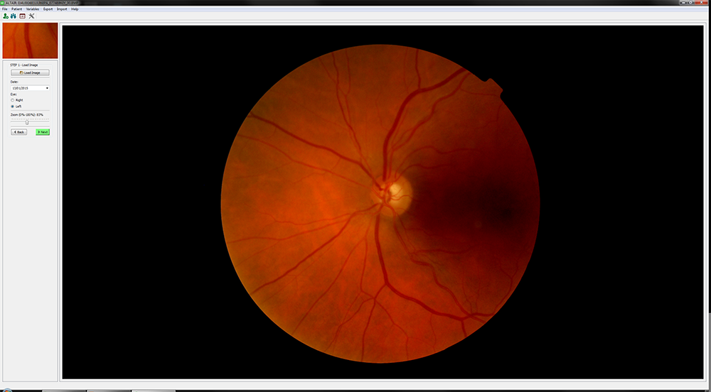
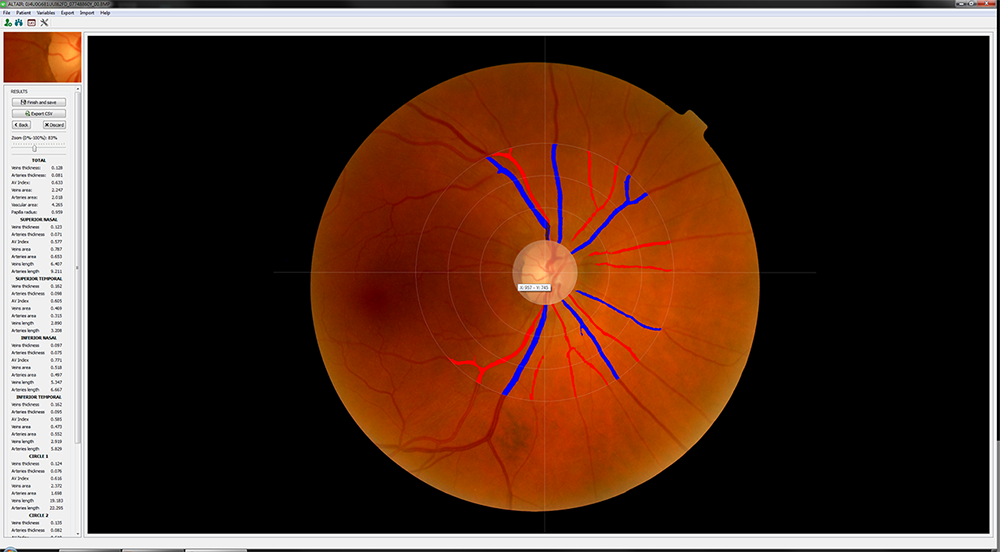
ALTAIR (Automatic image analyzer to assess retinal vessel caliber) is a novel platform image processing to study the structural properties of vessels, arteries and veins that are observed with a red-free fundus camera in the normal human eye, and the fractal analysis of the branching trees of the vascular system. Altair employs analytical methods and AI (Artificial Intelligence) algorithms to detect retinal parameters of interest. The sequence of algorithms represents a new methodology to determine the properties of retinal veins and arteries. The platform uses the latest computer techniques both statistical and medical:
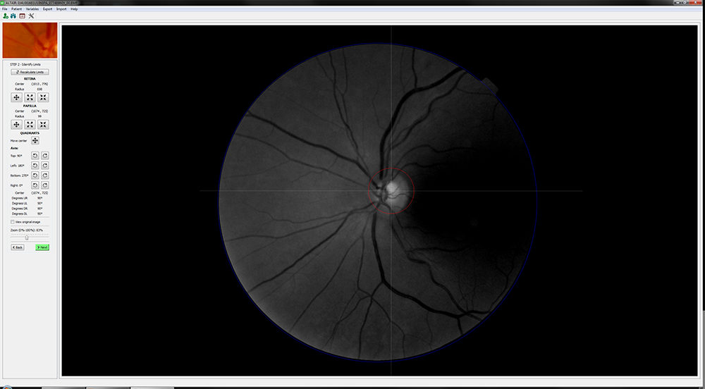
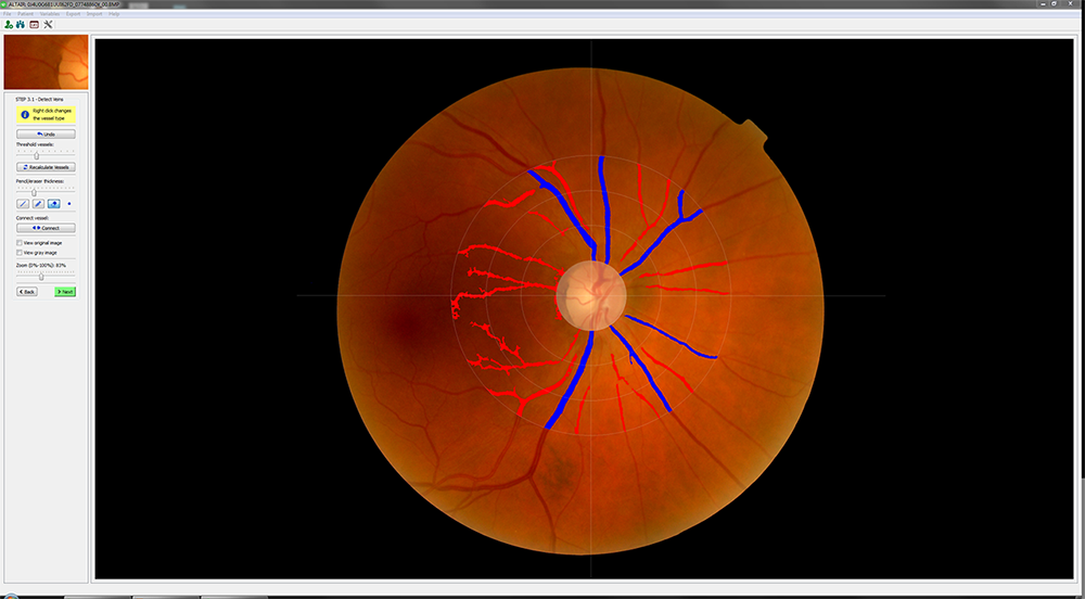
- Firstly, a phase called "digitization of the retina", in which the different parts of the eye image are identified. Here a data structure is created, which makes it possible to represent and process the retina without requiring the original image. This phase includes modules of preprocessing (techniques: filters noise, contrast, etc.) , detection (techniques based on the detection of edges or borders, Hough transform, etc.), and segmentation (algorithms based on matched filters, vessel tracking and Principal Component analysis or PCA).
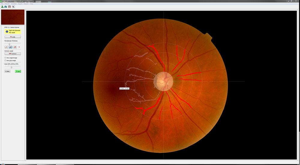
- Secondly, a phase of "measurements" in which we work with retinas that have been previously identified. This phase includes extraction of knowledge and manual correction (techniques: expert knowledge thresholding techniques, case-based reasoning (CBR), etc.).
The platform does not require user initialization, it is robust to the changes in the appearance of retinal fundus images typically encountered in clinical environments, and is intended as a unified tool to link all the methods needed to automate all processes of measurement on the retinas.
IMAGES